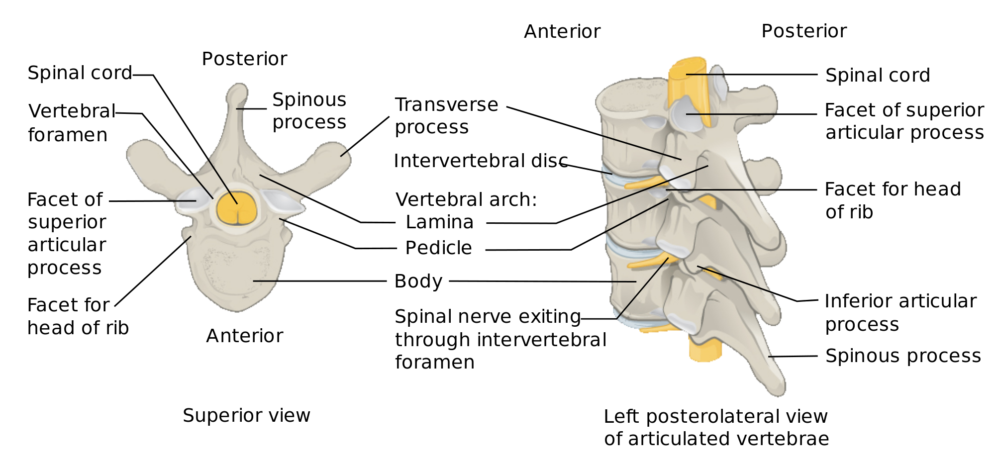
The sciatic nerve is formed by lumbar nerve roots (or as the picture below labels a “spinal nerve”) of L4,L5,S1,S2,and or S3. Some say that L3 is also involved. The differences of the lumbar nerve roots that make up the sciatic nerve depends not the researcher but I would not worry about that.
What you need to know is this, play close ATTENTION!
Depending on where the compression or pinching is occurring in the lumbar spine is where you will experience the numbness, tingling, weakness and or pain.Each nerve root has a specific area which provides sensation (aka dermatome) and or motor function (myotome) to certain muscles or parts of the leg, ankle, or foot. Let take for example if L4 nerve root is pinched by a herniate disc then ankle dorsiflexion is expected to be weak and the inner calf region is where it is expected for the numbness and or tingling to be expected compared to the not affected side. Remembers nothing in the body is exact so we are looking at the general pattern.
Here is a cheat sheet of general dermatome and myotomes of the lumbar nerve roots. Depending on the research, there may present with mild differences.